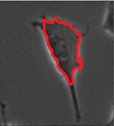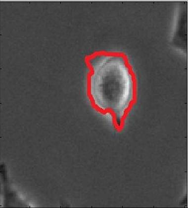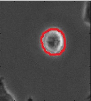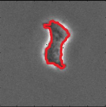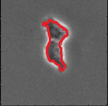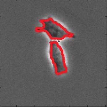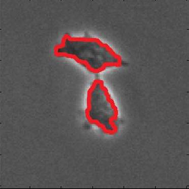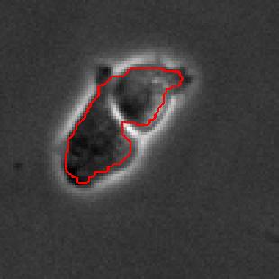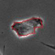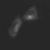

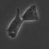

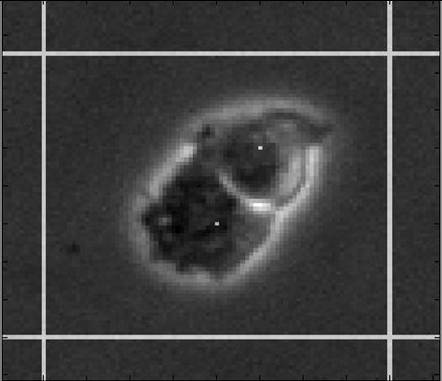
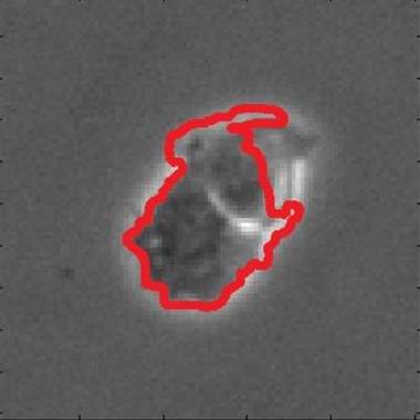
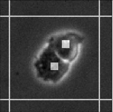
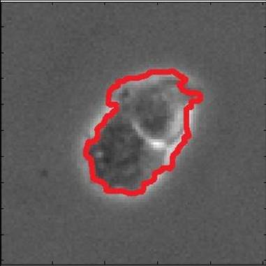
Segmentation of brightfield cell images from microscopy is challenging in several ways. The contrast between cells and the background is low. Cells are usually surrounded by ``halo'', an optical artifact common in brightfield images. Also, cell divisions occur frequently, which raises the issue of topological change to segmentation. In this research, we present a robust segmentation method based on the watershed and level set methods. Instead of heuristically locate where the initial markers for watershed should be, we apply a multiphase level set marker extraction to determine regions inside a cell. In contrast with the standard level set segmentation where only one level set function is used, we apply multiple level set functions (usually 3) to capture the different intensity levels in a cell image. This is particularly important to be able to distinguish regions of similar but different intensity levels in low contrast images. All the pixels obtained will be used as an initial marker for watershed. The region growing process of watershed will capture the rest of the cell until it hits the halo which serves as a ``wall'' to stop the expansion. By using these relatively large number of points as markers together with watershed, we show that the low contrast cell boundary can be captured correctly. Furthermore, we present a technique for watershed and level set to detect cell division automatically with no special human attention. Finally, we present segmentation results of C2C12 cells in brightfield images to illustrate the effectiveness of our method.


|  |  |  |
| bright field image | initial marker from level set | watershed segmentation | final result |