Jeff orchard’s research

My earlier research was in image processing, under the following categories.
Image Registration
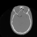
One of my favourite topics is medical image registration. I have been working on new cost functions to align images that were acquired with different modalities, such as aligning an MRI to a CT scan. I pay particular attention to speed. One method I developed uses the Fast Fourier Transform (FFT) to speed up the computation by orders of magnitude (50 to 500 times faster!).
Image Mosaics
An image mosaic is a rendering of a large target image by arranging a collection of small source images, often in an array, each chosen specifically to fit a particular block of the target image. I combine my registration methods with optimization techniques to produce such mosaics. Check out my cut-out image mosaics web page.
CT Reconstruction
My M.Math student Hwa-Young Kim (and previous M.Math student Alexei Ramotar) is working with me to develop reconstruction methods for next-generation portable CT scanners. Again, speed is a huge factor. The methods we are working on are very fast, and designed for a generation of portable CT scanners that aren't restricted to the circular geometry used in a hospital CT scanner.
Other
I have a general interest in image processing, including: Fractal Compression (downloads), Photomosaics, Photo-Stenciling.
( publications )
Computational neuroscience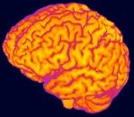
A normal human brain contains 85 billion neurons that pass electrical signals to each other. The brain exhibits macroscopic and microscopic organization. Somehow, this vast network of neurons takes sensory input (vision, hearing, etc.) and computes an appropriate response (motion, memories, etc.). But how do natural processes tune the neural connections so that animals - like humans - exhibit intelligent behaviour? What computational strategies does the brain use to do the amazing things it does?
In computational neuroscience, we model brain circuits using computer simulations of networks of spiking neurons. We build these models guided by neuroanatomy, and compare the results of the simulations to data collected from living brains. Usually, our models are coded into the Nengo neural simulation language.